HISTORY:
A male in his 30s presents with testicular pain. US scrotum shows vascular hypoechoic mass measuring 0.6 cm x 0.6 cm x 0.4 cm. Labs showed testosterone level of 225 ng/dl, AFP level of 1.2 ng/ml and beta-HCG level of <2.0 mIU/ml.
GROSS:
The specimen is designated "left testicle and spermatic cord; hx testicular cancer; s/p chemotherapy" and consists of a 53.8 gm orchiectomy specimen. The spermatic cord measures 10.1 cm in length x 2.1 cm in diameter. The testis measures 5.2 x 3.0 x 2.8 cm. The specimen is sectioned to reveal a tan-brown, firm nodule that measures 0.6 x 0.5 x 0.4 cm. The nodule measures 13.1 cm from the spermatic cord margin. The nodule is the same color as the remaining testicular parenchyma. The nodule does not involve the epididymis, rete testis, tunica albuginea or hilar fat. The remaining testicular parenchyma is tan-brown and strings with ease.
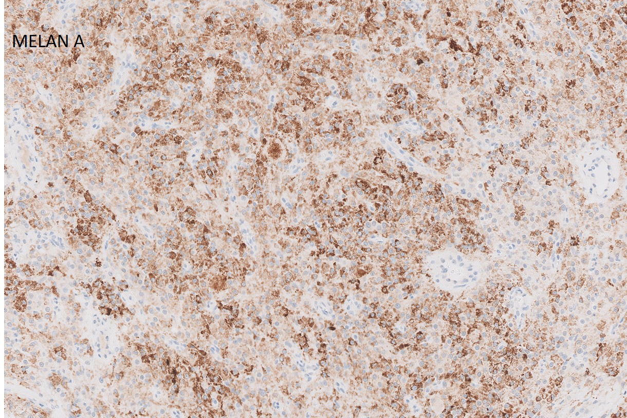
CD 117, Glypican 3 and beta-hcg stains were also done and read as negative.
What is the most likely diagnosis?
- Seminoma
- Leydig cell tumor
- Granular cell tumor
- Spermatocytic tumor
- Melanoma