Date of Presentation:3/4/2021

Attending pathologist: Anil Parwani, MD, PhD

Presented by: Bindu Challa, MD

Prepared by: Bindu Challa, MD

Organ:Kidney

HISTORY:

A male in his 50s presents with gross hematuria and flank pain. Computed tomography (CT) of abdomen and pelvis showed a 7 cm lobulated mass in right kidney.

GROSS:

Gross image of kidney
Microscopic image possibly detecting cancerous areas in human tissue

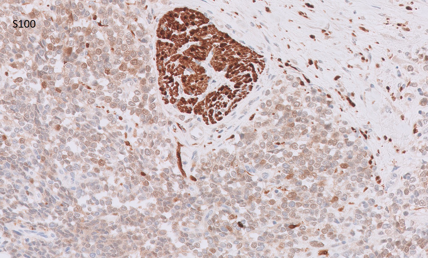
AE1/AE3, WT-2, CAIX, CD3, CD20, CD45, CK7 and CK20 stains were also done and read as negative.

What is the most likely diagnosis?

  1. Nephroblastoma
  2. Primary Ewing sarcoma of the kidney
  3. Desmoplastic small round cell tumor of kidney
  4. Lymphoma
  5. Small cell carcinoma of the kidney
Primary Ewing sarcoma of the kidney