N337C Doan Hall
410 W. 10th Ave
Columbus, OH 43210
Link to director's bio
Center of Excellence - Digital and Computational Pathology

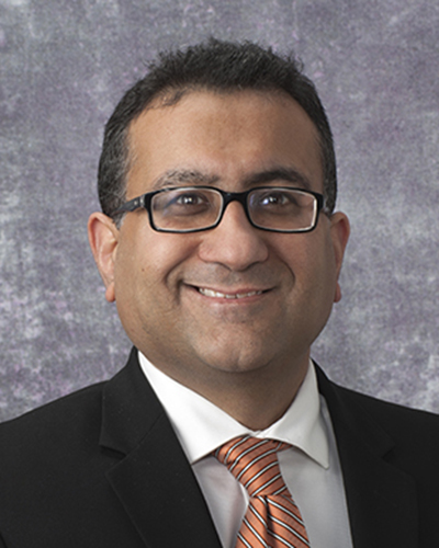
Giovanni Lujan, MD
Vice Chair, Clinical Informatics, Digital and Computational Pathology
Email
Email Dr. Lujan
The Ohio State University Wexner Medical Center is home to one of the world’s leading digital pathology programs.
Daily over 2,400 slides are digitized and made instantly available to our pathologists who use them for primary diagnosis, consultation, education, and research. In 2018, Ohio State became the first site in the United States to utilize digital pathology for primary diagnosis. Today, most of our sign-outs are digital.
Digital allows our pathologists to share their knowledge and skills to help patients around the state and across the globe. The use of digital images allows us to erase time and distance and provide pathology review in real time hundreds or even thousands of miles away.
We are proud to also be a leader in deploying quantitative image analysis (QIA), machine learning, and artificial intelligence to produce diagnoses for patients that are dramatically faster, more comprehensive, and more accurate than ever previously possible. We work with our partners to develop, test, and implement the tools that will transform pathology in the 21st Century.
Digital pathology has also enabled our teams to expand our research capabilities. Our research lab assists scientist with using digital resources and tools to probe the nature, causes, consequences, and treatment of disease in ways never before possible. Digital allows our researchers to assemble cohorts more rapidly, inquire of larger volumes of cases, and interrogate specimens more deeply, faster, and more accurately than ever before.
As a consequence of our endeavors, we manage one of the world’s largest collections of whole slide images (WSI). Ohio State is home to 4.7 million WSIs representing over 500,000 cases and growing, of every imaginable tissue and disease type.
Administration
Whole Slide Image (WSI) 20x - $16.00 - $30.00 depending on volume
Scanning of a 1" x 3" slide to create digital whole slide image at 20x magnification
Whole Slide Image (WSI) 40x - $16.00 - $30.00 depending on volume
Scanning of a 1" x 3" slide to create digital whole slide image at 20x magnification
Algorithm development analysis - $50.00+ per hour, dependent upon request
Creation of unique algorithm beyond simple measurement of a single feature
Image Analysis of Whole Slide (WSI) - $25.00+ per hour, dependent upon request
Quantification of features (region of interest size, nuclear staining, marker staining, nuclear size, etc.) for a digital image (non-TMA)
Slide analysis (20X / 40X) - $16.00 - $30.00 depending on volume
Slide Annotation - $16.00 - $30.00 depending on volume
Slide annotation/marking
Static Image (JPEG) - $16.00 - $30.00 depending on volume
Extraction of a JPEG image from a digital whole slide image at requested magnification (1x-20x)
TMA image analysis 100+ core - $75.00+ per hour, dependent upon request
Quantification of features (nuclear staining, marker staining, nuclear size, etc.) per core for a tissue microarray of more than 100 cores
TMA image analysis 1-100 core - $50.00+ per hour, dependent upon request
Quantification of features (nuclear staining, marker staining, nuclear size, etc.) per core for a tissue microarray of 100 or fewer cores

OSUCCC / OSU Pathology Scan Center
The OSUCCC – James Digital Pathology team of experts is dedicated to detecting and diagnosing cancer accurately and quickly. Our team’s mission is to work with researchers and physicians to provide answers to research questions—simple and complex—using the most sophisticated technology available.
The digital pathology process begins with whole-slide imaging, or scanning conventional glass slides and then using sophisticated technology to join consecutive images digitally into a single, whole image—replicating the information on the glass slide.
- (8) Philips Ultra Fast Scanners. Largest collection of such scanners in the country.
- Scan approximately 2500 slides a day
- Operational 24hrs a day / 5 days a week
- All slides are scanned at 40x magnification
- Each slide consist of apprximately 1GB of information
- Cancer Data Trust
Validation of digital pathology systems:
- CAP Validation completed for all 7 production Scanners for primary diagnosis scanning
- In progress: Whole mount Scanning for prostate and penile cancers
- Pdx External consults (since March 1): 782
- PDx cases (since March 1): 988
- Retrospective Cancer cases from thru Late 2013, 2014, 2015, 2016 & 2017 done and upto June 2018
- More than 720,000 Slides have been scanned
- More than 63,000 Cases are now available online
- First site in the US to go live with primary diagnosis
Adoption of digital pathology:
-
Tumor Boards & Conferences
- 37 Weeks of GYN tumor boards ( Used by Dr. Suarez, Dr. Li, Dr. Ivanov, Dr. Chen)
- Participated in MMC Breast tumor board in Spigelman center ( Dr. Tozbikian and Resident)
- Liver Conference ( Dr. Yearsley)
- Interstitial Liver Disease Conference ( Dr. Konstantin)
- Hem path ( Dr. Blower)
- Colorectal( Dr. Frankel)
-
Prospective External Secondary Consults
- ~ 690 cases have been digitized and read in parallel to glass slides
- Dr. Parwani, Dr. Li, Dr. Otero, Dr. Suarez & Dr. Konstantin are participating in the Pilot
-
Research IRB sets
- Endometrial cases library( Dr. Suarez and Dr. Li)
- Breast cases library ( Dr. Tozbikian)
- OCCPI
- Monthly QC, Daily QA digitized
- Daily Interesting Case : Starting Aug 2018
- Quality assurance slides digitized
- Send out slides digitized
- Research applications using visio pharm software
Selected Publications and Presentations
- Challa B, Kellough DA, Satturwar S, Lujan G, Frankel WL, Parwani AV, Sun S, Li Z. Detection of Lymph Node Metastasis of Breast Carcinomas Using Digital Imaging Analysis. United States & Canadian Academy of Pathology’s (USCAP) 111th Annual Meeting. Los Angeles, CA. March 19-24, 2022. (Poster)
- Vazzano J, Hu K, Johansson D, EurenK, Wilen LK, Zhou M, Parwani AV. INIFY Prostate* Predictions on Prostate Biopsies: Impact of Scanners, Preanalytical Factors and User Experience. United States & Canadian Academy of Pathology’s (USCAP) 111th Annual Meeting. Los Angeles, CA. March 19-24, 2022. (Poster)
- Jha S, Naik S, Baisakh M, Lobo A, Sharma S, Jain E, Balzer BL, Chakrabarti I, Parwani AV, Sahoo B, Mishra S, Sampat NY, Pattnaik N, Tiwari R, Mohanty SK. Solitary Fibrous Tumor (SFT) of the Adrenal Gland: A Multi-institutional Study of Nine Cases with Literature Review. United States & Canadian Academy of Pathology’s (USCAP) 111th Annual Meeting. Los Angeles, CA. March 19-24, 2022. (Poster#25 ; Abstract #112)
- Challa B, Shafi S, Parwani AV, Li Z. Digital Imaging Analysis of HER2 Immunohistochemistry in Endometrial Serous Carcinoma. United States & Canadian Academy of Pathology’s (USCAP) 111th Annual Meeting. Los Angeles, CA. March 19-24, 2022. (Poster)
- Kellough DA, Erck S, Palermini E, Li Z, Lujan, G, Shaker N, Parwani AV, Satturwar S. Types and Frequency of Whole Slide Imaging Scan Failures in a Clinical High Through-Put Digital Pathology Scanning Laboratory. United States & Canadian Academy of Pathology’s (USCAP) 111th Annual Meeting. Los Angeles, CA. March 19-24, 2022. (Poster)
- Kellough DA, Shafi S, Shaker N, Scarl R, Satturwar S, Lujan G, Frankel, W Li Z, Parwani AV. Drivers of Digital Pathology Adoption for Clinical Practice: An Experience from a Single Large Academic Institution. United States & Canadian Academy of Pathology’s (USCAP) 111th Annual Meeting. Los Angeles, CA. March 19-24, 2022. (Poster)
- Shafi S, Kellough DA, Lujan G, Satturwar S, Parwani AV, Li Z. Validation of Automated Digital Imaging Analysis of Estrogen Receptor Immunohistochemistry in the Clinical Setting. United States & Canadian Academy of Pathology’s (USCAP) 111th Annual Meeting. Los Angeles, CA. March 19-24, 2022. (Poster)
- Lujan G, Kellough DA, Frankel WL, Chhibber NN, Parwani AV, Chen W, Li Z, Lombardo A, Walsh J, Lloyd M. Faster Than Glass: A Digital Pathology Workflow Unlocks Major Time-Savings. United States & Canadian Academy of Pathology’s (USCAP) 111th Annual Meeting. Los Angeles, CA. March 19-24, 2022. (Poster)
- Vazzano J, Parwani AV, Johansson D, Euren K, Wilen LK, Zhou M. INIFYProstate* predictions on prostate biopsy: Impact of preanalytical factors and user experience. Pathology Visions 2021. Las Vegas, NV, October 17-19, 2021. (Poster)
- Hamasaki S, Nohle DG, Ayers LW, Parwani AV. Do tissue pre-analytical and analytical factors influence the performance of tissue morphometrics/AI machine learning algorithms for tumor quantification? OSU Center for Clinical and Translational Science (CCTS) 2019 Annual Scientific Meeting, The Ohio State University, Columbus, OH, December 3, 2019. Poster. (1st Place Poster – Staff)
- Li AC, Yearsley K, Hammond S, Li Z, Parwani AV. Quantification of estrogen receptor immunohistochemistry using whole slide imaging and morphometric image analysis software. College of American Pathologists (CAP) Annual Meeting, Orlando, FL. Sep 12-15, 2019. Published abstract in Arch Pathol Lab Med 2019 Sep;143(9):e224-e225. Poster 166.
- Lloyd M, Cuddihy M, Shulgay T, Curtiss C, Krueger G, Mack D, Pietryka A, Gill T, Parwani AV. Sectioning automation to improve quality and decrease costs for a high-throughput slide scanning facility. United States & Canadian Academy of Pathology (USCAP) 108th Annual Meeting, National Harbor, MD, Mar 16-21, 2019. Published abstract in Mod Pathol, 2019 Mar;32(3 Suppl):35-36 and Lab Invest, 2019 Mar;99(3 Suppl):35-36. Abstract 1937.
Note: Click on heading text to expand or collapse.

